AI-powered zebrafish phenotyping
Point at a frame to see what the models find in it.
How it reads an embryo
Start with a plate. Pick a well.
Each well holds a zebrafish embryo exposed to a compound. Pick any well.
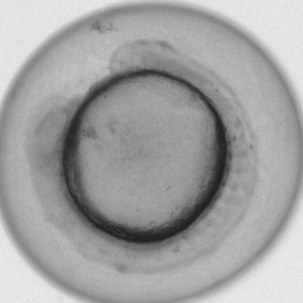
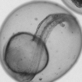
MorphoScan images every well on a schedule. Each well holds a living zebrafish embryo that was exposed to a compound. Pick any well. They are real embryos from the EmbryoNet paper's test sets, and the authors checked every label. Whichever you choose takes the same route through all three models.
The plate is an illustration: 48 real frames arranged like one.
Two models, one image.
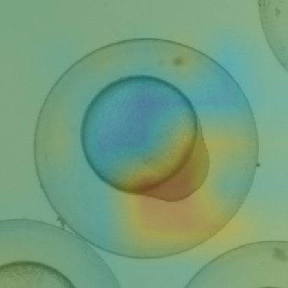
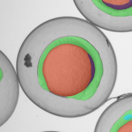
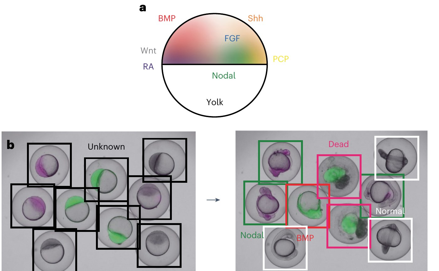
The Segmenter outlines the tissues. The Classifier names the pathway and marks the regions behind its call.
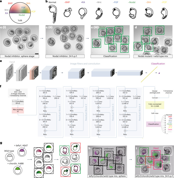
The Segmenter (MorphoNet) colors in the tissues, pixel by pixel. The Classifier (EmbryoNet) takes in the whole embryo, says which pathway the phenotype points to, and highlights the parts of the image that led to its call.
How old is it?
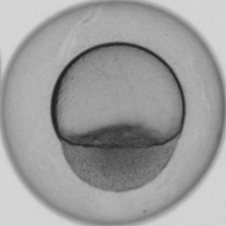
The Stager compares the embryo with reference embryos of every age and takes the closest match.
Each frame is resized to the scale of the reference images first.
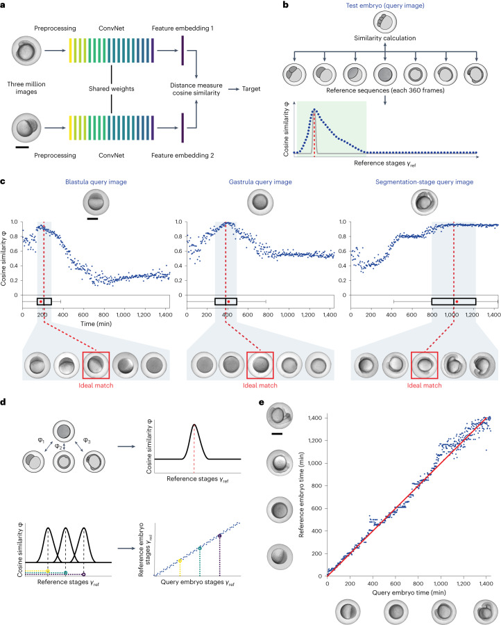
The Stager (TwinNet) doesn't use a clock. It compares the embryo with real reference embryos of every age and takes the age of the closest one. Watch the frame find its match.
Before matching, each frame is resized to the scale of the reference images.
Image in, mechanism out.
Run any model on every frame of a plate and export the results.
Run any of the three models on every frame of a plate, not one embryo at a time, and export the tissue maps, the ages or the pathway calls.
See the modelsPlatform
Capture with MorphoScan. Analyze in the app.
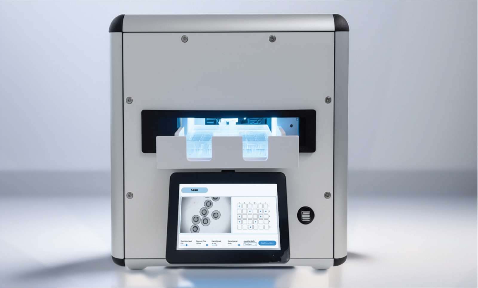
A high-throughput microscope for standard laboratory incubators.
- Fits inside your incubator.
- Captures images on a schedule.
- Designed for longitudinal imaging of embryos and organoids.
- Not tied to one species. It images whatever is in the plate.
Analyze the images in the web app
All of these are live today. More species are coming on top of them, and more models are in development.
Science
Peer-reviewed before it was a product.
The methods behind Morphomics come from the Müller Lab at the University of Konstanz, and they featured in Nature Methods' Method of the Year 2023.
Team
Morphomics brings MorphoScan, EmbryoNet, MorphoNet and TwinNet together. It grows out of EmbryoNet AI Technologies and the Müller Lab at the University of Konstanz.
Contact
Run a pilot.
Whether you're in pharma, at a CRO, in a tox lab or in an academic group, we can send you a MorphoScan for your own samples, or analyze the images you already have. We are also open to collaborations: bring your data and your questions, and we will build new models with you.