Real data, real models
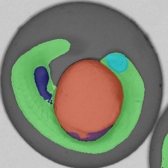
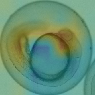
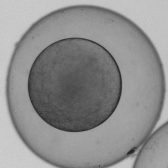
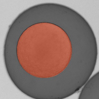
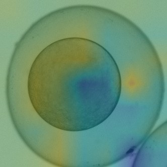
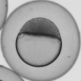
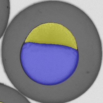
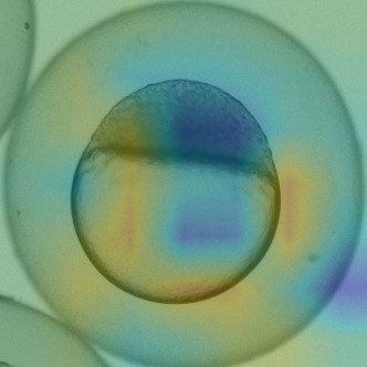
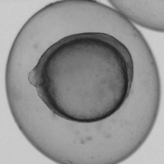
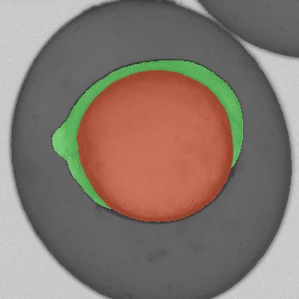
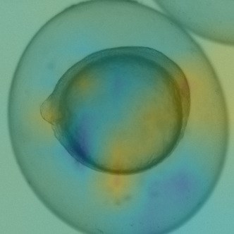
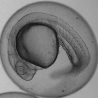
What you see is unprocessed. Point at one to see what we detect.
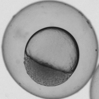
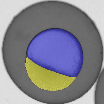
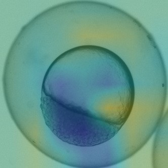
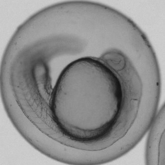
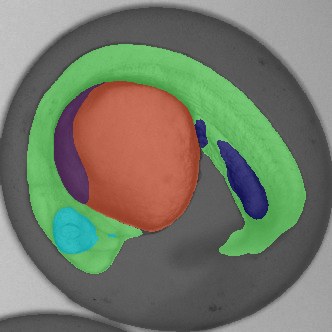
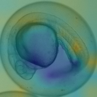
Real data, real models
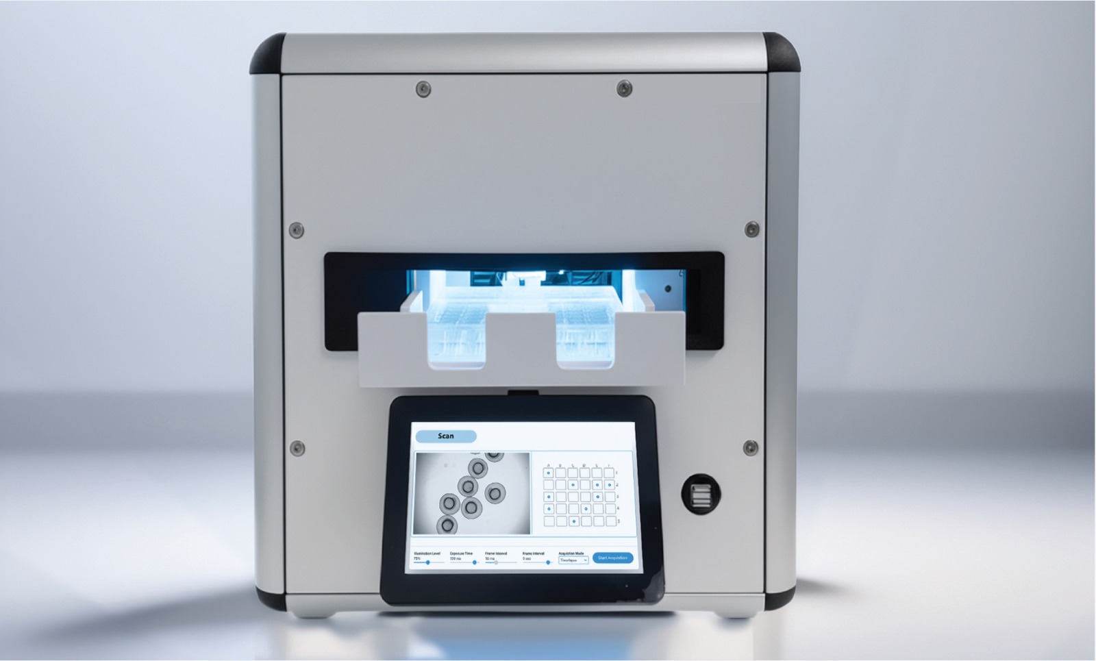
MorphoScan images embryos and organoids inside a standard incubator. Four AI modules turn what it records into quantitative phenotype data.
Module 1 — Classifier
Module 2 — Segmentor
Module 3 — Stager
Module 4 — Explorer
MorphoScan images embryos and organoids inside a standard incubator. Four AI modules turn what it records into quantitative phenotype data.
Module 1 — Classifier
Module 2 — Segmentor
Module 3 — Stager
Module 4 — Explorer
Technology
Drug discovery, toxicology, and developmental research depend on image-based biological models to understand how chemical, genetic, and environmental perturbations affect living systems. When development goes wrong, the damage is written into the body's shape — a trained biologist can look at a malformed embryo and tell which signalling pathway was disrupted.
Reading that phenotype remains one of the most direct ways to find out what a substance does to a living organism. Extracting it at scale is the bottleneck: manual evaluation is labour-intensive, hard to standardise between evaluators, and the high-content imaging platforms that automate it demand infrastructure and expertise most labs don't have.
Morphomics addresses this with an integrated platform — MorphoScan, a compact automated imaging system that sits inside a standard incubator and records time-lapse images of embryos and organoids, combined with four AI modules that transform those images into quantitative phenotypic information.
Pricing
MorphoScan instrument
€25,000
One-time hardware purchase. Sits inside your existing incubator — no dedicated imaging room required.
Starter
€299/mo
Analysis volume for roughly one full 48-well plate per month.
Professional
€699/mo
Analysis volume for roughly three full 48-well plates per month.
Multi-site
€1,199/mo
Analysis volume for roughly six full 48-well plates per month, across sites.
A full 48-well plate produces around 20,000 images. Additional analysis volume beyond a tier's allowance is available through usage-based top-up packages.
Publications
The methods behind Morphomics originated in the Müller Lab at the University of Konstanz and were recognised as part of Nature Methods' Method of the Year 2023.
Method of the Year 2023
EmbryoNet: using deep learning to link embryonic phenotypes to signaling pathways
Read paper →Method of the Year 2023
Uncovering developmental time and tempo using deep learning
Read paper →Method of the Year 2023
AI identifies developmental defects and drug mechanisms in embryos
Read paper →More publications will be added here.
About us
Science & Hardware
AI models and MorphoScan's imaging hardware.
Software, AI & Deployment
The hosted platform, inference, and model registry.
Commercial & Financing
Pricing, go-to-market, and commercial strategy.
Scientific Mentor
Chair of Developmental Biology, University of Konstanz.
Contact
Academic lab, biotech, or CRO — get in touch and we'll help you evaluate MorphoScan for your screening workflow.
hello@morphomics.de